Our Office
5909 South Congress Ave
Lake Worth, FL 33462
Existing Patients: (561) 967-6453
New Patients: (561) 203-0359
Emergency: (754) 800-1949
Fax: (561) 431-5866
Visit Us Online

Digital radiography utilizes computer technology and digital sensors for the acquisition, viewing, storage, and sharing of radiographic images. It offers several advantages over the older traditional film based methods of taking x-rays. The most significant of these advantages is that digital radiography reduces a patient’s exposure to radiation. Other benefits are that images can be viewed instantly after being taken, can be seen simultaneously as needed by multiple practitioners, and can be easily shared with other offices. Digital x-rays are also safer for the environment as they do not require any chemicals or paper to develop.
An electronic pad, known as a sensor is used instead of film to acquire a digital image. After the image is taken, it goes directly into the patient’s file on the computer. Once it is stored on the computer, it can be easily viewed on a screen, shared, or printed out.
Digital radiography uses computer technology and electronic sensors to capture diagnostic images of teeth, bone and surrounding oral structures. Instead of traditional film, a small sensor records x-ray data and transfers it directly to a computer, where the image is stored in the patient record. This workflow supports faster diagnosis, easier recordkeeping and immediate review of images.
Because images appear instantly on-screen, clinicians can adjust contrast, magnification and orientation to evaluate detail without re-exposing the patient. Digital files are simple to archive and share with other providers when clinical coordination is needed. The process is also more environmentally friendly since it eliminates film and chemical processing.
Digital radiography replaces photographic film with an electronic sensor and computer software, producing images instantly without chemical development. The technology streamlines the imaging process because clinicians can view, enhance and store images immediately at the chairside. Software tools allow for image enhancement that can make subtle findings easier to detect compared with film.
Another important difference is integration with electronic records, which reduces physical storage needs and improves access to prior images. Digital systems also tend to reduce the number of repeat exposures because clinicians can confirm image quality on the spot. Taken together, these differences speed appointments and improve clinical communication.
Digital radiographs are considered safe when taken according to accepted dental guidelines and exposure-minimization principles. Sensors generally require less radiation than film to produce diagnostic images, and size-appropriate techniques further reduce dose for pediatric patients. Protective measures such as lead aprons and thyroid collars are used as standard practice to limit exposure.
For patients who are pregnant or may be pregnant, dental teams follow established precautions and image only when clinically necessary, positioning shielding appropriately. The decision to proceed with imaging balances diagnostic benefit and minimal risk, and clinicians will discuss timing and alternatives with the patient. Clear communication helps ensure necessary care is provided safely and with patient comfort in mind.
Digital sensors are more sensitive to x-rays than film, so clinicians can obtain high-quality diagnostic images using lower exposure settings. Image-enhancement software reduces the likelihood of retakes by correcting contrast and brightness without additional radiation. Immediate review of images allows technicians to reposition or adjust technique on the spot when necessary.
Practices also combine sensor efficiency with standard radiation safety measures such as proper collimation, optimized exposure settings and the use of protective aprons. These measures follow the principle of keeping radiation as low as reasonably achievable while still obtaining the images needed for accurate diagnosis. The overall approach reduces cumulative exposure for patients over time.
Digital radiographs provide detailed views of tooth roots, bone levels, interproximal decay and other structures that are critical for diagnosis. Clinicians use these images to detect cavities, evaluate periodontal bone loss, assess root morphology prior to endodontic treatment and plan restorations or extractions. The ability to magnify and adjust images enhances detection of subtle conditions that inform treatment decisions.
For treatment planning, digital images integrate easily with clinical photos and intraoral scans to create comprehensive records that guide restorative, orthodontic and surgical procedures. Images can be annotated and added to the patient chart to illustrate findings and explain options during consultations. This integrated documentation supports predictable treatment outcomes and efficient care coordination.
Yes. One of the major advantages of digital radiography is the ability to export and share image files quickly with specialists, referring dentists and laboratories. Digital formats preserve diagnostic detail and allow consultants to review cases in advance, which can streamline referrals and treatment planning. Rapid sharing reduces the need for duplicate imaging and speeds care coordination.
When images are transmitted outside the practice, they are sent using secure methods and only to authorized recipients for the purpose of patient care. Electronic transfer preserves image quality and enables efficient collaboration among providers. This capability improves continuity of care and helps ensure timely clinical decisions.
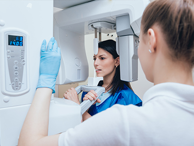
During a digital radiography appointment a small sensor will be placed inside the mouth or a panoramic unit will be positioned depending on the type of image required. The actual x-ray exposure is brief and painless, and most patients only notice the presence of the sensor during positioning. The clinician will review the image immediately and confirm whether additional views are needed.
If more images are necessary adjustments can be made quickly without a long delay, often avoiding the need for a separate appointment. Chairside monitors allow the dentist to show and explain findings directly to the patient, which helps with understanding and informed decision-making. Patients with anxiety or special needs should inform the team so positioning and communication can be adapted for comfort.
Digital images are treated as part of the protected health record and are stored on access-controlled systems with routine backups to prevent loss. Office protocols typically include user authentication, role-based access and staff training to maintain confidentiality and limit access to authorized personnel. Regular software updates and secure network practices help reduce the risk of unauthorized access.
When images are shared with outside providers they are transmitted through encrypted channels and only to recipients involved in the patient's care. The practice maintains documentation and policies for handling health information to support consistent privacy protections. Patients may request secure copies of their images and the office will provide them after appropriate verification.
Digital radiography shortens appointment times by eliminating film processing and enabling immediate image review, which reduces waiting and streamlines diagnosis. Enhanced imaging tools can reveal early-stage problems that are easier to treat, improving preventive care and long-term outcomes. Clear, on-screen visuals also help clinicians explain conditions and treatment options in language patients can understand.
Because digital images are archived and easily retrievable clinicians can compare current and prior views to monitor changes over time and adjust care as needed. Faster sharing with specialists reduces delays in referrals and treatment planning, which can contribute to better clinical results. Overall, the technology supports more efficient, transparent and patient-centered care.
The practice follows manufacturer recommendations and scheduled maintenance for sensors, x-ray generators and imaging software to preserve consistent image quality. Routine quality assurance checks include calibration, sensor inspection and software updates so that diagnostic images remain reliable. Staff receive ongoing training in positioning and equipment handling to minimize artifacts and the need for retakes.
In addition to equipment upkeep, the team adheres to radiation safety protocols such as proper collimation, exposure settings and protective barriers to safeguard patients and staff. Records of maintenance and safety checks are kept as part of continuous quality improvement and clinical governance. These measures help deliver dependable imaging while minimizing risk.