Our Office
5909 South Congress Ave
Lake Worth, FL 33462
Existing Patients: (561) 967-6453
New Patients: (561) 203-0359
Emergency: (754) 800-1949
Fax: (561) 431-5866
Visit Us Online

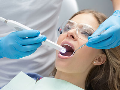
An intraoral camera is a small, pen-sized imaging device that captures high-resolution, color images from inside the mouth and displays them instantly on a monitor. Because the camera can be positioned close to a tooth or soft tissue, it reveals fine details—hairline cracks, early decay, worn restorations, and subtle soft-tissue changes—that might be difficult to see with the naked eye alone. For patients, those live images translate complex findings into something clear and understandable.
Designed to be comfortable and noninvasive, the intraoral camera slips easily into the mouth and is maneuvered precisely by a clinician. Its compact tip and focused lighting allow the device to photograph occlusal surfaces, interproximal areas, and the gum line without prolonged discomfort. The result is a visual record that supports faster, more confident clinical observations during routine exams and targeted evaluations alike.
When used thoughtfully, this technology becomes more than a camera: it’s a diagnostic extension of the clinician’s senses. It complements traditional visual exam techniques and digital X-rays by providing a true-color surface view that helps both clinicians and patients see what’s happening in real time.
High-quality intraoral images assist clinicians in detecting early signs of disease and planning appropriate care. Magnified views can reveal fractures, defective margins on crowns and fillings, and enamel defects before they become symptomatic. These images help the clinician document progression over time and determine whether a conservative approach or a restorative intervention is warranted.
Intraoral cameras also support more precise treatment planning. When a potential problem is clearly documented, clinicians can select targeted interventions—such as a focused restoration, sealant placement, or periodontal therapy—with greater confidence. The images can be reviewed alongside radiographs and other diagnostic data to form a complete, evidence-based treatment strategy.
Beyond diagnosis, consistent imaging improves continuity of care. Clear photos in the patient record make it easier to monitor healing after a procedure or to compare changes at follow-up visits, reducing uncertainty and helping clinicians track outcomes objectively.
One of the most valuable aspects of the intraoral camera is its ability to create shared understanding. When patients see an enlarged image of their own tooth or gum tissue, complex explanations become tangible. Viewing an image together fosters informed conversations about oral health, treatment options, and preventive measures so patients can play a more active role in decisions about their care.
Sharing images in real time also helps set realistic expectations. Patients can see the exact condition of a restoration, the margin of a crown, or the early-stage discoloration on a tooth, which makes recommendations more transparent and easier to accept. This visual clarity supports collaborative care and strengthens the clinician–patient relationship.
In addition to clinical conversations, intraoral imagery can be an effective teaching tool. Hygienists and clinicians can point out plaque accumulation, explain brushing and flossing techniques in context, and demonstrate how simple behavior changes can affect specific areas of the mouth.
Intraoral camera images are fully digital and integrate cleanly into contemporary clinical workflows. Captured photos can be stored directly in the patient’s chart, organized alongside radiographs and written notes, and retrieved later for comparison. This digital continuity simplifies charting and supports thorough clinical documentation.
When collaboration with outside specialists or dental laboratories is needed, annotated images can be shared to convey clinical details that are difficult to describe in words alone. Photographic records help ensure that laboratory partners and consulting clinicians receive a consistent, accurate representation of the clinical situation, which improves coordination and efficiency.
Patient privacy and data security are important considerations with any electronic record. Images acquired with intraoral cameras are handled according to established privacy standards and stored within secure practice systems, ensuring that visual records remain part of a confidential clinical file rather than public content.
An intraoral camera exam is straightforward and typically takes only a few minutes as part of a regular checkup or a focused evaluation. The clinician will gently position the camera to capture specific teeth or soft-tissue areas, often while the patient watches the live image on a monitor. The process is noninvasive and generally more comfortable than some other diagnostic procedures.
Captured images are reviewed immediately and may be annotated or saved for future reference. If a concern is identified, the clinician will explain the findings using the image as a visual aid, outline the recommended next steps, and discuss how the images will be used in the patient’s ongoing care. If monitoring is appropriate, the practice will compare future images to these initial photos to track any changes.
For families and patients seeking clarity about oral health, the intraoral camera is a reassuring tool: it reduces guesswork, documents clinical observations, and supports shared decision-making. At Horizon Palms Family Dentistry in Lake Worth, this technology is one of several modern tools used to make exams more informative and treatment discussions more productive.
To summarize, intraoral cameras bring immediate, detailed visual information to the dental exam—enhancing diagnosis, improving communication, and supporting integrated digital records. If you’d like to learn more about how intraoral imaging is used during visits or how it may benefit your care, please contact Horizon Palms Family Dentistry for more information.
An intraoral camera is a small, pen-sized imaging device that captures high-resolution, color photographs from inside the mouth. The clinician positions the camera close to teeth or soft tissues and the device transmits live images to a monitor for immediate review. Because it provides a true-color surface view with focused lighting, the camera reveals fine details that may be difficult to see with the naked eye.
The camera’s compact tip and magnification allow clinicians to document occlusal surfaces, interproximal areas, and the gum line with precision. Images are captured digitally and can be saved into the patient record for later comparison. This technology serves as a visual extension of the clinical exam and complements radiographs and clinical palpation.
High-quality intraoral images help clinicians detect early signs of disease such as hairline cracks, marginal gaps in restorations, enamel defects, and subtle soft-tissue changes. Magnified photos make it easier to determine whether a conservative approach or a restorative intervention is warranted. These images are reviewed alongside radiographs and clinical findings to create an evidence-based treatment plan.
Consistent photographic documentation also enables clinicians to monitor progression over time and evaluate healing after procedures. Clear images reduce diagnostic uncertainty by providing objective visual evidence that can be reexamined during follow-up visits. This leads to more targeted treatment decisions and improved continuity of care.
An intraoral camera exam is typically quick and noninvasive, often taking only a few minutes as part of a routine checkup or focused evaluation. The clinician will gently maneuver the camera while the patient may watch live images on a monitor, which can help explain findings in real time. Captured photos may be annotated or saved to the chart for future reference.
If a concern is identified, the clinician will use the image to explain the issue, describe recommended next steps, and outline monitoring plans if appropriate. Patients are encouraged to ask questions while viewing the images to better understand their oral health. At Horizon Palms Family Dentistry in Lake Worth these images are used to support clear, informed discussions during the visit.
Intraoral cameras are designed to be comfortable and noninvasive, with a small, rounded tip and focused lighting to minimize discomfort. The device is maneuvered gently and does not emit ionizing radiation, so it poses no radiation risk unlike X-rays. Most patients find the procedure easier and less intrusive than some other diagnostic methods.
The camera’s safety profile makes it appropriate for patients across age groups, including children and older adults, provided they can tolerate a short intraoral examination. Clinicians always take care to work slowly and explain each step to reduce anxiety. Standard infection-control protocols are followed to ensure the device is clean and safe for each use.
Intraoral camera images are fully digital and are saved directly into the patient’s electronic chart alongside radiographs and clinical notes. Images can be organized, annotated, and retrieved later for comparison, which simplifies charting and supports thorough documentation. Digital integration streamlines clinical workflows and makes it easier to assemble a complete record of a patient’s oral health.
When multiple clinicians or team members are involved in care, shared access to annotated photos improves communication and consistency. Photographic records also assist in tracking treatment outcomes and documenting healing or disease progression. Secure practice systems ensure images remain part of the confidential patient file rather than public content.
Yes, intraoral images are often shared with specialists and dental laboratories to convey clinical details that are difficult to describe in text alone. Annotated photos help outside providers understand the exact condition of a tooth, restoration margin, or soft-tissue concern and can improve coordination of care. Sharing images supports clearer instructions for laboratory work, such as shade selection or restoration design.
When images are transmitted to outside providers, practices use secure methods consistent with privacy standards to protect patient information. Clear photographic documentation reduces the need for repeated explanation and can speed collaborative treatment planning. This contributes to more predictable outcomes and efficient case management.
Viewing enlarged, true-color images of their own teeth and gums helps patients understand clinical findings that might otherwise seem abstract. Shared images turn complex explanations into a tangible, visual conversation, enabling patients to make more informed decisions about preventive measures and treatment options. This shared review often leads to clearer expectations and greater confidence in care plans.
In addition to diagnosis, intraoral imagery is a practical teaching aid for hygiene instruction and behavior change. Clinicians and hygienists can point out plaque accumulation or demonstrate brushing and flossing techniques on the actual images, making guidance more actionable. Patients who see the specific areas that need attention are often better equipped to follow targeted home-care recommendations.
While intraoral cameras excel at showing surface details and true-color images, they do not replace radiographs, tactile examination, or other diagnostic tools needed to evaluate underlying structures. Cameras cannot visualize bone, deep interproximal decay under intact enamel, or pathology hidden beneath restorations. Clinicians use intraoral images as one component of a comprehensive diagnostic process rather than a standalone solution.
Image quality can be affected by patient movement, limited mouth opening, or lighting conditions, which may require additional imaging or clinical assessment. Proper interpretation requires professional training to avoid over- or underestimating clinical significance. When limitations are present, clinicians combine camera images with other diagnostic data to reach accurate conclusions.
Intraoral images are stored within a practice’s secure electronic health record system and handled according to applicable privacy regulations and internal policies. Access controls, encrypted storage, and secure transmission methods help ensure that photographic records remain confidential and are only available to authorized personnel. Proper labeling and organization within the chart maintain clear documentation for future reference.
When images must be shared with other providers for consults or laboratory collaboration, secure transfer protocols are used to protect patient information. Patients may be informed about how their images will be used for diagnosis, treatment planning, and continuity of care. Horizon Palms Family Dentistry follows standard privacy practices to keep visual records part of a protected clinical file.
The frequency of intraoral imaging varies depending on clinical need; images are commonly captured during regular exams, targeted evaluations, and pre- or post-treatment visits. Clinicians may repeat photos to monitor suspicious areas, document healing, or compare changes over time. Capturing images at baseline and at follow-up visits provides an objective record to assess progression or resolution of a condition.
Repeated imaging supports evidence-based decisions about when to intervene and helps track the effectiveness of conservative treatments or periodontal therapy. Clinicians will determine the appropriate schedule based on the individual patient’s risk factors and clinical findings. Consistent photographic documentation enhances long-term monitoring and continuity of care.